A new mouse MRI atlas been added to BrainGlobe#
Dorr et al. (2008) created a high-resolution, three-dimensional MRI-based atlas of the adult C57Bl/6J mouse brain, providing comprehensive anatomical coverage of the cerebrum, cerebellum, and brainstem.
The atlas was constructed from within-skull, T2-weighted MR images acquired at 32 μm isotropic resolution from forty 12-week-old mice scanned at 7T. Following normalisation, registration, and averaging of individual scans, 62 distinct brain structures were manually delineated.
This atlas is now available through BrainGlobe as dorr_mouse_mri_32um.
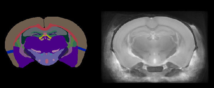
Figure 1. Two-dimensional view of the Dorr mouse MRI brain atlas: annotations (left) and reference (right).
How do I use the new atlas?#
You can use the Dorr mouse MRI atlas like all other BrainGlobe atlases. To visualise the atlas, you could follow the steps below:
Install BrainGlobe (instructions).
Open Napari and follow the steps in our download tutorial for the
dorr_mouse_mri_32umatlas.Run
napari -w brainrender-napariand visualise the different parts of the atlas as described in our visualisation tutorial.
The end result will look something like Figure 2.
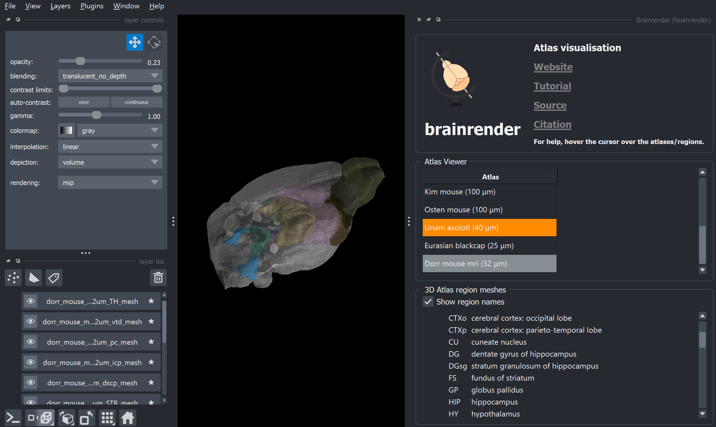
Figure 2: The Dorr mouse MRI atlas visualised with brainrender-napari.
Why are we adding new atlases?#
A fundamental aim of the BrainGlobe project is to make various brain atlases easily accessible by users across the globe. If you would like to get involved with a similar project, please get in touch.